


ENT Imaging
Our research group, in collaboration with our partners at the University Hospital for Otorhinolaryngology, Head and Neck Surgery, focuses on various aspects of imaging and image analysis.
One primary focus is the application of flat-panel detectors, cone-beam computed tomography (CBCT), and photon-counting CT in temporal bone imaging (Figs. 1–2), as well as the use of artificial intelligence for virtually enhancing the spatial resolution of standard CT datasets of the temporal bone.
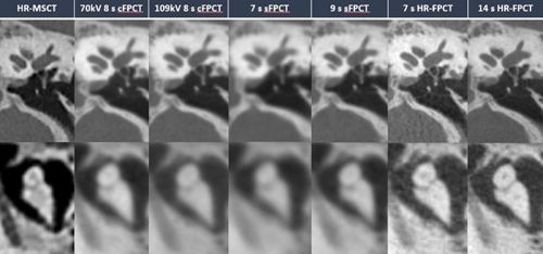
Figure 1
Illustrative example of the different protocols in the visualization of middle ear landmarks, stapes (top row) and incudomalleolar joint (lower row) Only HR-FPCT protocols allowed for superior visualization of the stapedial landmarks and obvious delineation of incudostapedial joint.
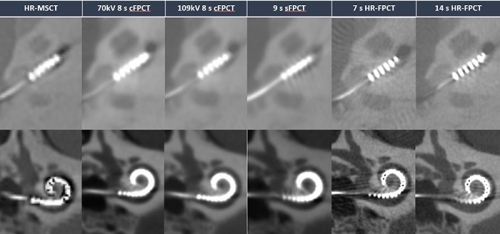
Figure 2
Illustrative example of different protocols in the evaluation of electrode array, axial view (top row) and paracoronar view (lower row) of the cochlea Only HR-FPCT protocols allowed for superior discernability of individual electrode contacts and inter-contact spaces.
Another key area of interest is the establishment and investigation of standardized reporting for common ENT examinations in neuroradiology, including CT and MRI of the neck, CT of the paranasal sinuses, and CT of the temporal bone. Additionally, in collaboration with the Chair of Visualization (Prof. Dr.-Ing. Bernhard Preim), we are developing virtual reality (VR) applications (Fig. 3) for educational purposes in ENT and neuroradiology.
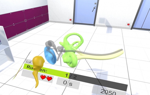
Figure 3
Screenshot taken from our current gamified VR teaching tool for middle and inner ear anatomy.
Video Project Ear
The video shows a part of our gamiefied teaching tool for ear anaytomy in 3D and correlation to temporal bone CT images
Group Members:
Doctoral Candidates:
Cooperation Partners:
University Clinic for Otorhinolaryngology, Head and Neck Surgery:
Computer Science (OVGU):
Computer Science (Kiel):
Publications
|
Diamandis E, Müller S, Khadhraoui E, Klebingat S, Einspänner E, Durisin M, Albrecht A, Behme D. Accelerated Flat Panel Computed Tomography for Pre-operative Temporal Bone Imaging: Image Quality and Dosimetry Comparison to Conventional High Resolution Multislice Computed Tomography. Neuroradiology 2025 (in press) |
|
Diamandis E, Müller S, Khadhraoui E, Klebingat S, Durisin M, Albrecht A, Behme D. Cochlear Implant Imaging with Accelerated Flat-Panel CT: Image Quality and Dosimetry Comparison to Conventional High-Resolution Multislice CT. The Neuroradiology Journal 2025 (in press) |
|
Tim Härtel, Mareen Allgaier, Daniel Behme, Bernhard Preim, Sylvia Saalfeld |
|
Sudden Hearing Loss and Vertigo After Sinus Occlusion. |